6 Single and multislice helical CT scan[32]. Download Scientific Diagram
Spiral Computed Tomography. Spiral computed tomography is a type of 3D CT scan. A CT scan (or CAT scan) is a non-invasive diagnostic imaging procedure that uses a combination of special X-ray equipment and sophisticated computer technology to produce cross-sectional images (often called slices), both horizontally and vertically, of the body.
Aquilion PRIME CT Scanner Technology Computed Tomography Canon Medical Systems USA
Helical CT (also called spiral CT) has several advantages over older CT techniques: it is faster and produces better quality 3-D pictures of areas inside the body, which may improve detection of small abnormalities. CT has many uses in the diagnosis, treatment, and monitoring of cancer, including. screening for cancer.

Helical CT for the Evaluation of Acute Pulmonary Embolism AJR
A low-dose helical CT scan is a quick, painless exam that takes multiple 3-dimensional pictures of the chest moving in a spiral motion around the body. As compared to a traditional CT scan, a low-dose CT scan produces five times less radiation.
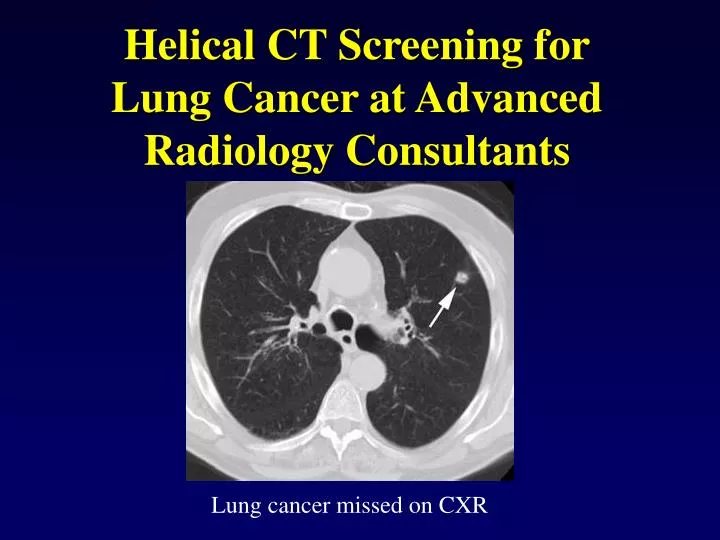
PPT Helical CT Screening for Lung Cancer at Advanced Radiology Consultants PowerPoint
However, helical CT requires one to be more cognizant of the relationship between contrast material administration and scanning, since the optimal temporal window for detection of disease can be missed. Factors unique to helical technology can produce artifacts, which one must be aware of when interpreting helically generated scans.
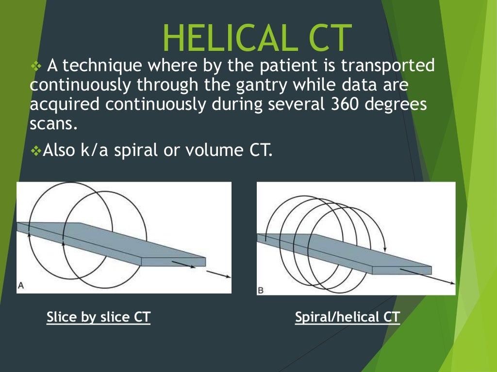
Helical and Multislice CT
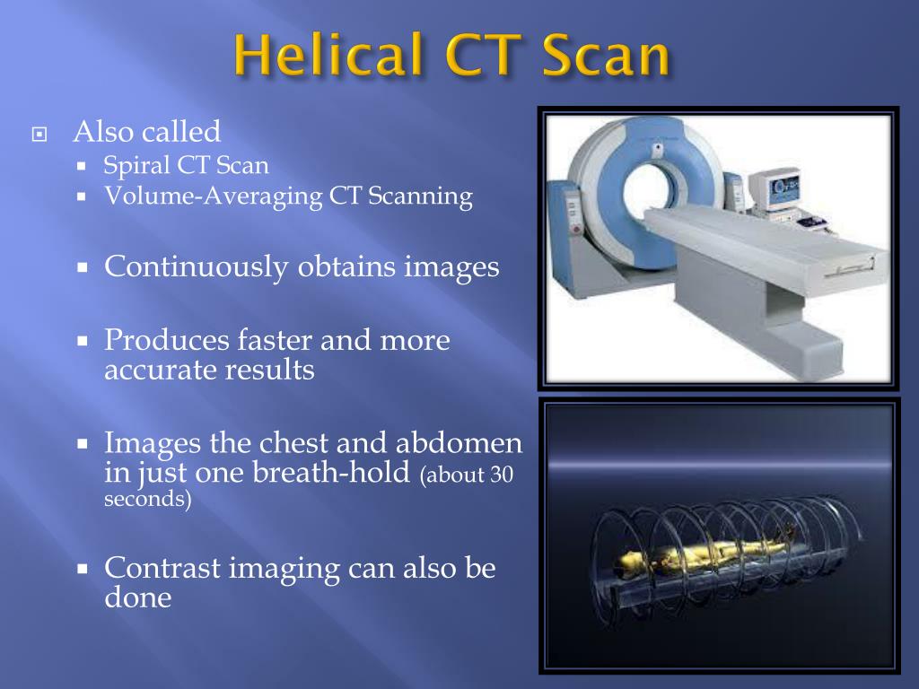
Helical (Spiral) CT is a vast improvement over conventional CT scans. The patient lies on an exam table that passes through a doughnut-shaped scanner, while an X-ray tube rotates around the table. This movement results in a spiral shaped continuous data set without any gaps. With the helical CT, there is less likelihood to miss small tumors or.
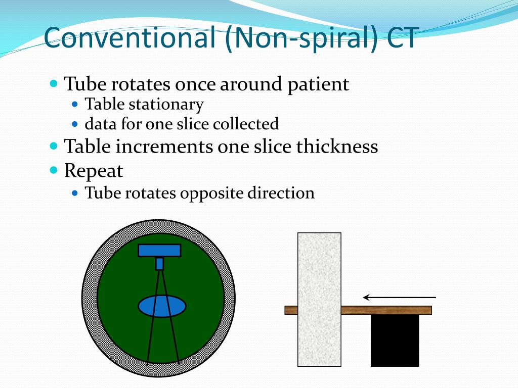
PPT Seeram Chapter 13 Single Slice Spiral Helical CT PowerPoint Presentation ID5515829
Spiral (helical) computed tomography (CT) involves continuous patient translation during x-ray source rotation and data acquisition. As a result, a volume data set is obtained in a relatively short period of time. For chest or abdominal scanning, an entire examination can be completed in a single breath hold of the patient or in several.
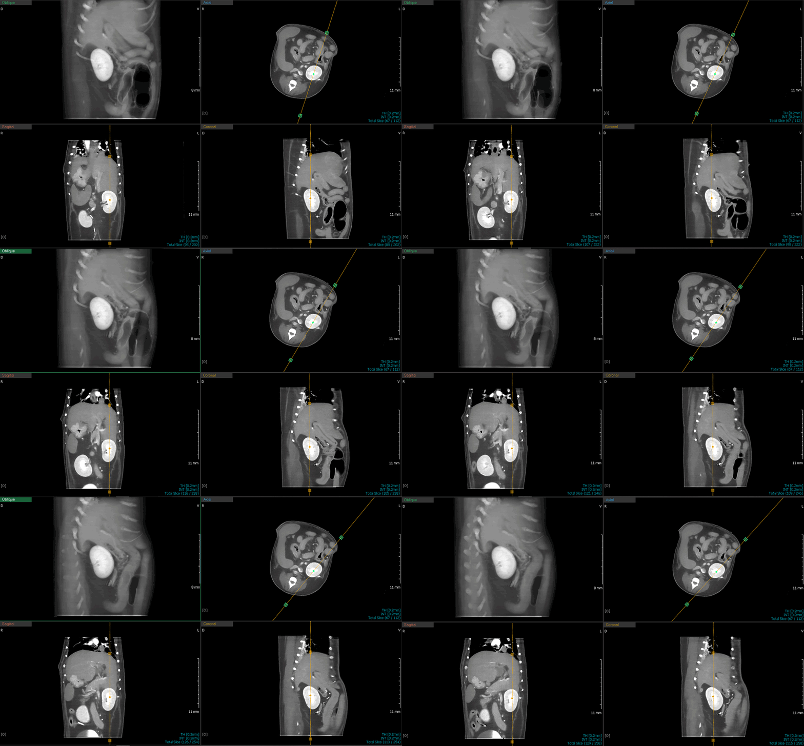
HelicalLinear CT Scanner Helical CT Scanner woorien
Slip-ring CT is the technology which enables power and data to be transferred without physical cables connecting the stationary and rotating portions of the CT gantry. The slip-ring enables continuous gantry rotation which in tern enables multiple acquisition types including: helical CT, cine CT, cardiac CT, CT perfusion, gated lung imaging (i.

16 Slice Helical Medical CT Scanner/ Medical Computed Tomography Scanning Machine MSLCT16 R
Helical (spiral) computed tomography (CT) is having a dramatic impact on body imaging. Unlike conventional CT, helical CT provides continued volumetric acquisition as the patient moves through the gantry. Advantages of helical CT include dramatically shortened examination times, improved visibility of vascular structures, better enhancement of parenchymal organs, the capability for.

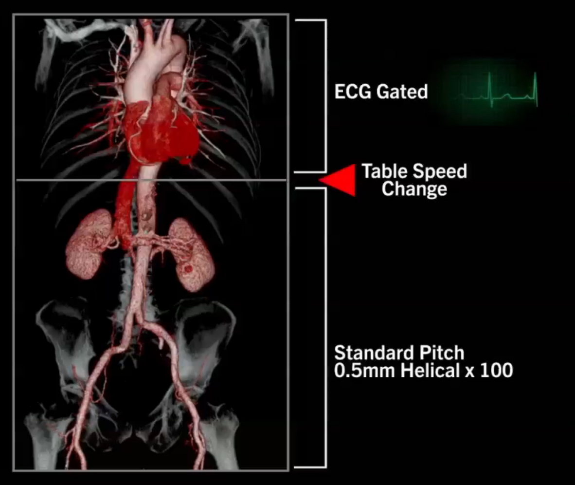
SPIN RADIOGRAPHERS Helical CT Evaluation of the Thoracic Aorta Helical CT Aortography Technique
Spiral computed tomography, or helical computed tomography, is a computed tomography (CT) technology in which the source and detector travel along a helical path relative to the object. Typical implementations involve moving the patient couch through the bore of the scanner whilst the gantry rotates.
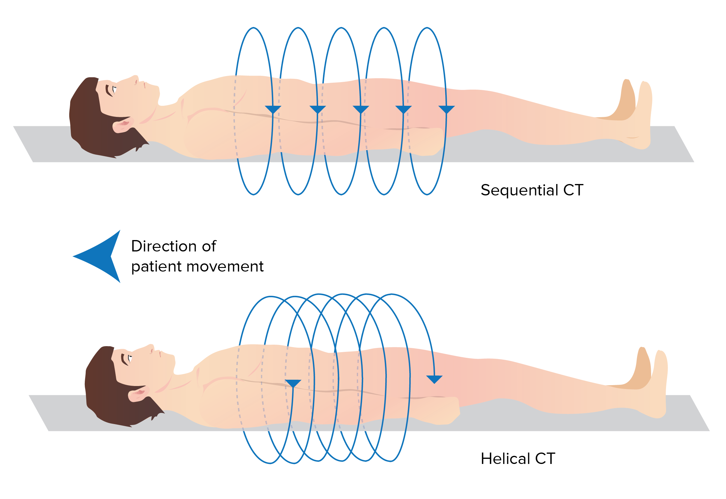
Computed Tomography (CT) Concise Medical Knowledge
The performance of helical CT requires several user-defined parameters that exceed the requirements of conventional CT. One needs to carefully select the collimation, table increment, and reconstruction interval. Minimizing these parameters maximizes longitudinal resolution but with various trade-offs. Decreasing the collimation decreases the effective section thickness but increases pixel.
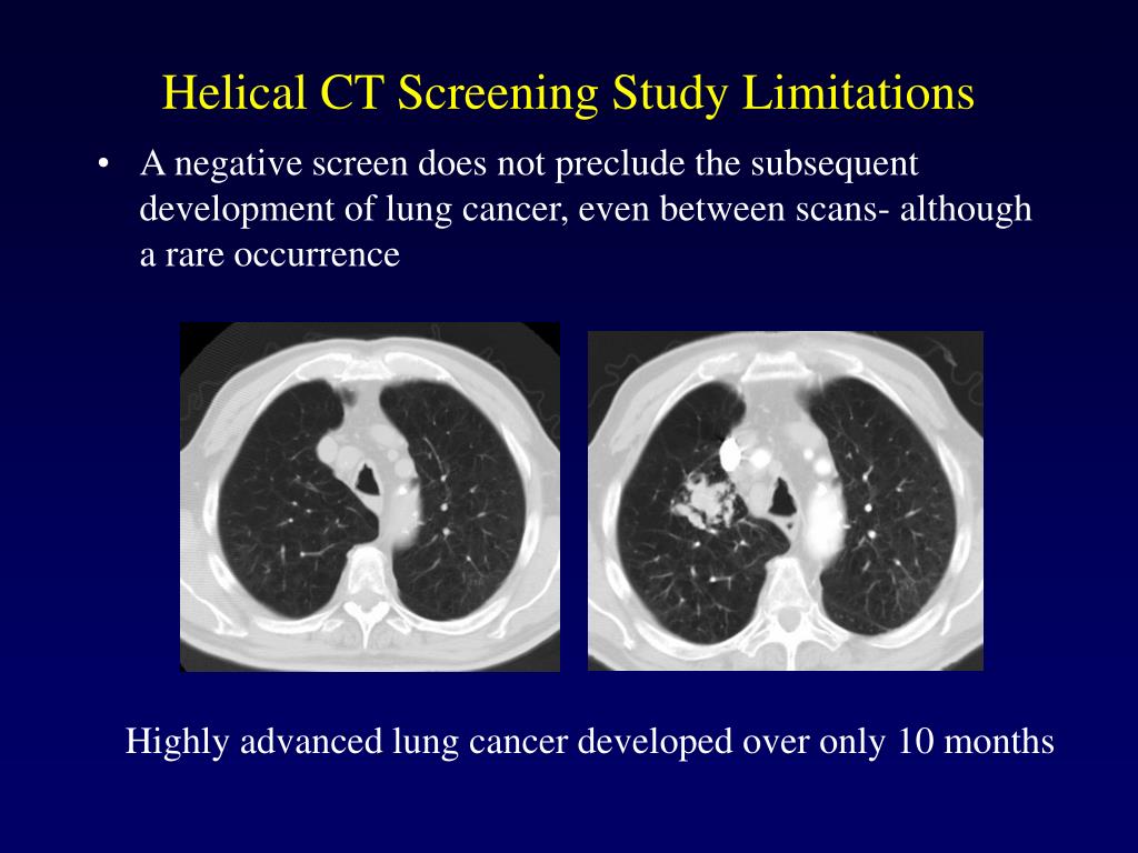
PPT Helical CT Screening for Lung Cancer at Advanced Radiology Consultants PowerPoint
For typical CT scanners the table is moving during the scan, as this is equivalent to the source and detector assembly moving in the other direction during the scan. In helical scanning an important descriptor of the acquisition is the helical pitch.

Dualphase helical CT, transverse scan on the level of the superior... Download Scientific Diagram
Helical (a.k.a. spiral) CT image acquisition was a major advance on the earlier stepwise ("stop and shoot") method. With helical CT, the patient is moved through a rotating x-ray beam and detector set. From the perspective of the patient, the x-ray beam from the CT traces a helical path. The helical path results in a three-dimensional data set.
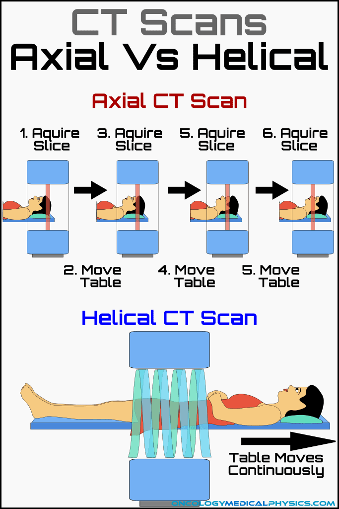
CT Design and Operation Oncology Medical Physics
The performance of helical CT requires several user-defined parameters that exceed the requirements of conventional CT. One needs to carefully select the collimation, table increment, and reconstruction interval. Minimizing these parameters maximizes longitudinal resolution but with various trade-offs. Decreasing the collimation decreases the.

Axial Vs Helical CT Scan
Helical CT is also useful in revealing vascular complications such as pseudoaneurysms and splenic and portal vein thrombosis [98, 101]. Fig. 16. —57-year-old man with acute necrotizing pancreatitis and severe back pain. CT scan using scan protocol III shows large region of unenhancement (necrosis) involving most of body and tail of pancreas.

3.6 Computed Tomography (CT) Medicine LibreTexts
A CT scan is a diagnostic imaging procedure that uses a combination of X-rays and computer technology to produce images of the inside of the body. It shows detailed images of any part of the body, including the bones, muscles, fat, organs and blood vessels. CT scans are more detailed than standard X-rays. In standard X-rays, a beam of energy is.
PPT PN 132 Day 2 PowerPoint Presentation, free download ID2258506
Spiral (helical) computed tomography (CT) involves continuous patient translation during x-ray source rotation and data acquisition. As a result, a volume data set is obtained in a relatively short period of time. For chest or abdominal scanning, an entire examination can be completed in a single breath hold of the patient or in several successive short breath holds. The data volume may be.
- James Cook Hotel Grand Chancellor Nz
- Native Animals In Western Australia
- What Word Rhymes With Word
- The Day The Crayons Quit Book
- Who Is Lesbian In The Matildas
- What Is A First Amendment Auditor
- How Are Plastic Bottles Made
- 720 X 2040 Internal Door
- Pacific Championship 2023 Team List
- 24 Hr Blood Pressure Monitoring
