

EMRad Radiologic Approach to the Traumatic Ankle
Standard X-rays are performed for many reasons, including diagnosing tumors or bone injuries. X-rays are made by using external radiation to produce images of the body, its organs, and other internal structures for diagnostic purposes. X-rays pass through body structures onto specially-treated plates (similar to camera film) and a "negative.
Ankle Sprains My Family Physio
For self-care of an ankle sprain, use the R.I.C.E. approach for the first two or three days: Rest. Avoid activities that cause pain, swelling or discomfort. Ice. Use an ice pack or ice slush bath immediately for 15 to 20 minutes and repeat every two to three hours while you're awake.
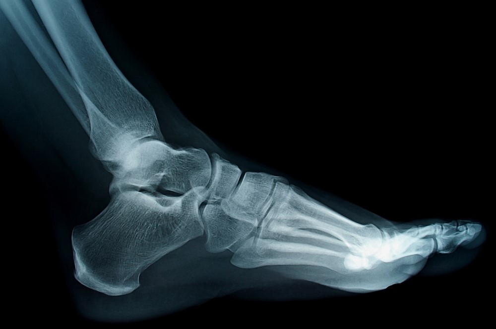
Healthy ankle joint xray of a 79 year old female patient. No fracture nor sprain can be seen
Simply put, there are 5 reasons to get an ankle x-ray. 1. You can't walk on the ankle. 2. You have tenderness at the outside ankle bone (fibula). 3. You have tenderness at the inside ankle bone (tibia). 4. You have tenderness at the outside of the foot (at the "styloid").

Sprained Ankle X Ray
Sprained Ankle. Ankle sprains are common injuries that occur among people of all ages and at all activity levels; in fact, they are the number one reason for missed participation in athletics. An ankle sprain occurs when the strong ligaments that support the ankle stretch beyond their limits and tear. The severity of a sprain can vary greatly.

Broken or Sprained Ankle? Pontchartrain Orthopedics & Sports Med
It's time to get an X-ray if your child has any of the following symptoms: Tenderness over the bony prominence on either side of the ankle, particularly if it's in the back. Inability to bear any weight for four steps (limping counts as weight-bearing). Tenderness over the outer or inner edge of the foot (where the foot is widest).

Ankle xrays
The ankle is a synovial joint composed of the distal tibia and fibula as they articulate with the talus. The distal tibia and fibula articulate with each other at the distal tibiofibular joint which is more commonly referred to as the tibiofibular syndesmosis (or simply the syndesmosis). As with all films, check around each bone on the film.
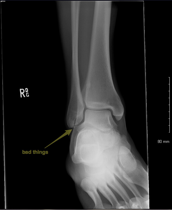
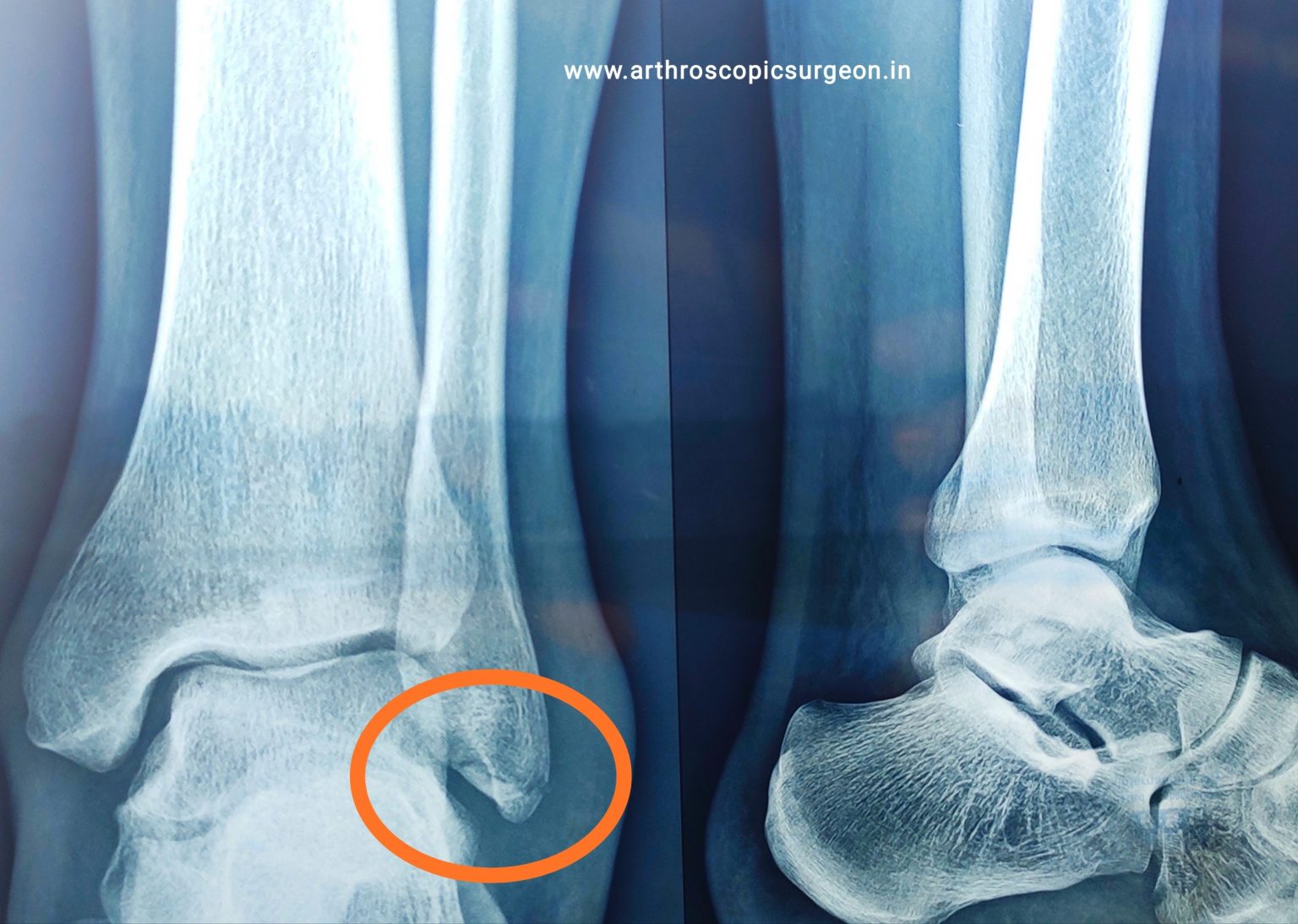
ankle xray fibula chip fracture(s) and low ankle sprain Mike Matz Flickr
Normally, after 2-3 weeks you will have decreased range of movement, strength and balance from your ankle injury. Here are some key rehab exercises to start with: Calf stretch: 1-2 minutes, 2-3 times a day. Keep the back knee straight to feel a stretch! Calf raises: 8-10 reps, 2-3 times a day. Start with two feet and aim for equal weight.

XRay of ankle I sprained my ankle in France this summer, … Flickr
Sudden pain or pulling in your ankle when you twist or injure it. A popping sound from your ankle when you twist or injure it. Pain or tenderness when touching your ankle. Swelling. Bruising. Inability to bear weight on the affected leg. Your doctor may recommend a splint or support while the sprain heals.
Sprained Ankle Your Questions Answered rugbystore Blog
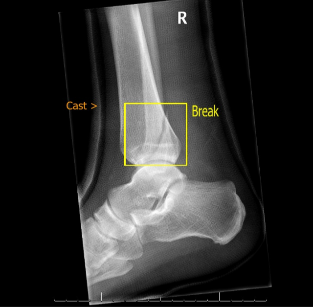
A broken ankle or ankle fracture is when one or more of the bones in your ankle break. Severe sprains and fractures have similar symptoms (pain, swelling, bruising, tenderness) and are both caused by twisting or rotating your ankle, tripping or falling, or trauma to your ankle. Sprains heal faster, but it can take up to six weeks for a broken.

X Ray Of Sprained Ankle
Inversion Ankle Sprains. The most common type of ankle sprain occurs when the foot is inverted, twisting inwards. When this type of ankle sprain happens, the outer, or lateral, ligaments are stretched too far. There are three lateral ankle ligaments that support the outer side of the joint. About 90% of ankle sprains are inversion injuries.

High Ankle Sprain X Ray
Getting medical attention right away if you sprain your ankle is vital. Especially true if you have severe pain, swelling, bruises, or trouble putting weight on your foot. A doctor or nurse may order an X-ray to rule out multiple fractures. If a sprain is found, it is usually treated with rest, ice, compression, and elevation, which all help to.
Ankle sprain Best Orthopedic doctor in Jaipur
Emergency rooms and urgent care facilities will perform X-rays. Specifically, a 5th metatarsal base is a common fracture in ankle sprains. If this occurs, wear a knee-high cam walking boot and crutches. These fractures are very slow to heal and often take up to three months. Surgery may be required if the fracture is displaced or separated.

Investigating the Validity of Soft Tissue Signs on Lateral Ankle XRay to Aid Diagnosis of
Sprained ankle vs. broken ankle. While a sprained ankle involves torn or stretched ligaments, a broken ankle means that at least one of the bones in your ankle is broken. Symptoms of a sprain and.

Ankle Fractures Core EM
Ankle sprains are a common sports-related injury and account for 5% of all emergency department visits. There is a high probability of a fracture If a patient meets the criteria of Ottawa's rules. However, if no fracture is evident on x-ray, then extra-articular swelling directs the clinician to the diagnosis of an ankle sprain since ligaments.

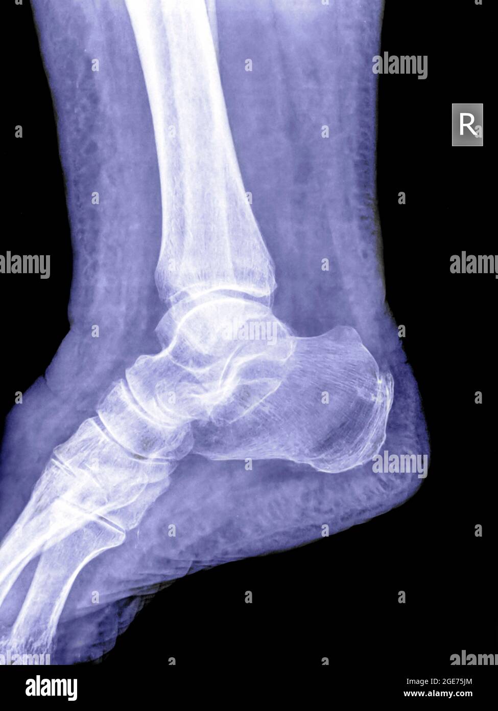
(A) Anteroposterior radiograph of the left ankle joint of a 22yearold... Download Scientific
The most common ankle injury is ankle sprain and of ankle sprains, a lateral ankle sprain is by far the most common, accounting for up 75-80% of ankle sprains 1. There is a higher incidence in children and adolescents than in adults 2,3. Women are thought to be more frequently affected 2,4.

Xray Picture Of A Sprained Ankle Stock Photo Download Image Now Analyzing, Anatomy, Ankle
Acute ankle sprains are commonly seen in both primary care and sports medicine practices as well as emergency departments and can result in significant short-term morbidity, recurrent injuries, and functional instability. Although nonoperative treatment is often successful in achieving satisfactory outcomes, correct diagnosis and treatment is important at the time of initial evaluation to.
- Did John Ibrahim Ever Go To Jail
- Australian World War Two Medals
- Stars V Renegades Scorecard Today
- Mount Isa To Gold Coast Flights
- Can You Defend Yourself In Australia
- Law And Order Svu Fanfiction
- How Much Is A Perm In Australia
- Hoka Women S Clifton 8 Stores
- Bull Mastiff Pitbull Mix Puppy
- South Island Of New Zealand Map