
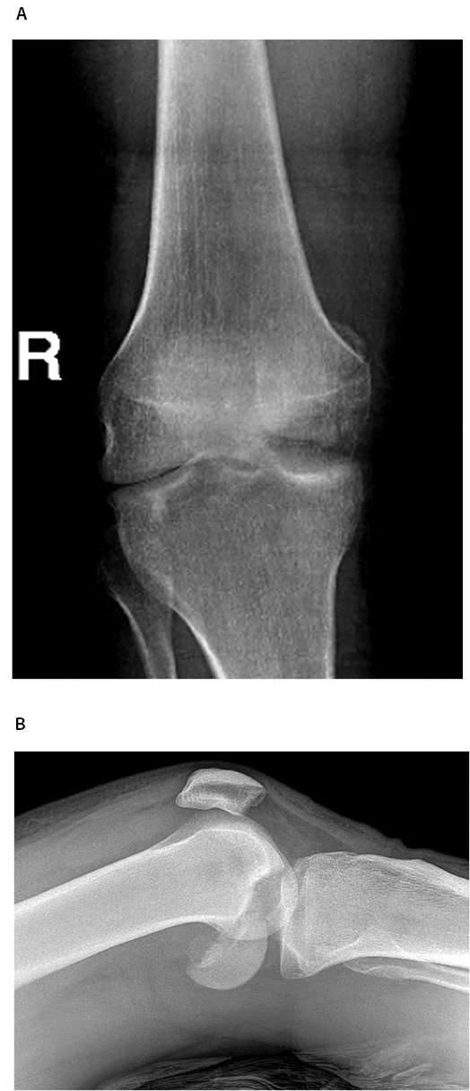
Medial Femoral Condyle Fracture
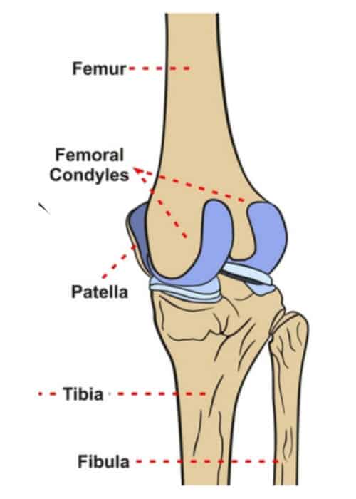
The femur is a long bone that widens at its distal end, these flared parts are called the medial and lateral condyles. Fracture of femoral condyle can occur, although it is a rare injury. It accounts for only about 5% of fracture to the femur, and that is less than 0.5% of all fractures.

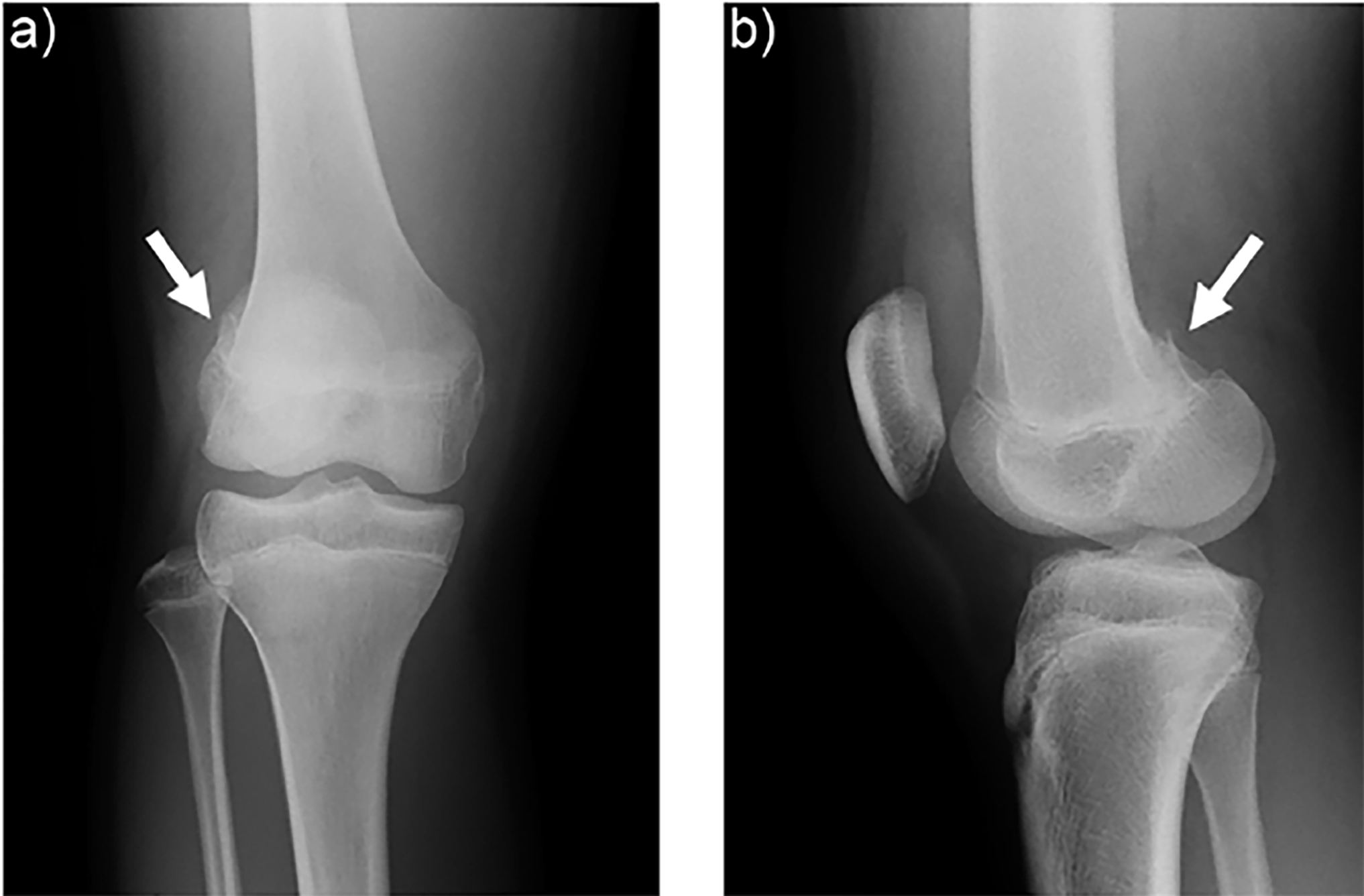
Cureus A Case of Distal Femur Medial Condyle Hoffa Type II(C) Fracture Treated with Headless
femoral condyle or patella/trochlear groove. Microfracture technique is a reparative technique used to treat articular cartilage defects. It is indicated for full-thickness articular cartilage loss in a weightbearing area between the femur and tibia or between the patella and trochlear groove.
Medial Femoral Condyle Fracture Fixation Images
Distal femoral fractures involve the femoral condyles and the metaphyseal region, commonly caused by high energy trauma such as motor vehicle accidents or a fall from a height. In the elderly, they may occur from falling at home. Other names: Supracondylar femur fracture; Intercondylar femur fracture; Hoffa's Fracture. The majority of distal femur fractures require surgical intervention.

Cureus A Case of Distal Femur Medial Condyle Hoffa Type II(C) Fracture Treated with Headless
A high-energy injury resulting in a Hoffa fracture of the medial condyle is often associated with a tibia fracture, a bicondylar Hoffa fracture, [44,45] a dislocation of the patella, a knee dislocation, intercondylar and supracondylar fractures, [9,47] and pelvic [48,49] and femoral shaft fractures.
Distal Femur Fracture TeachMeSurgery
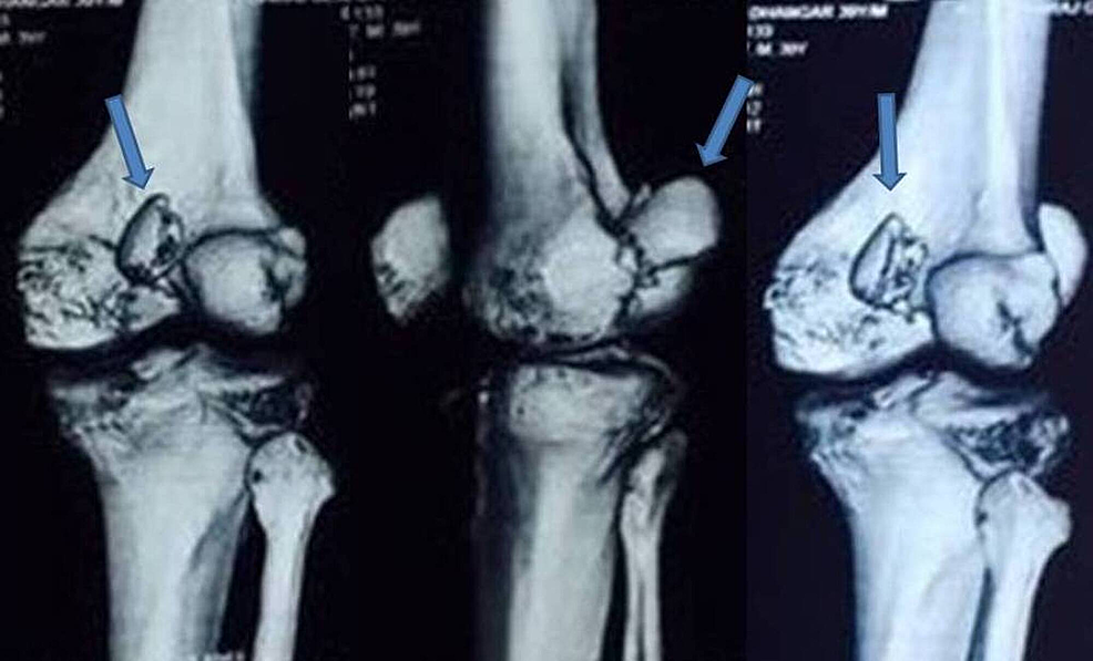
CT is extremely useful in diagnosing the presence of a "Hoffa" fragment, a coronal plane fracture, most commonly involving the lateral femoral condyle. 5 Temporizing measures typically involve immobilization with a long-leg splint or knee immobilizer. In cases of extreme deformity, a closed reduction may be required.

Cureus A Case of Distal Femur Medial Condyle Hoffa Type II(C) Fracture Treated with Headless
Medial • Useful for isolated medial condyle fractures or severely comminuted fractures in which medial fixation is required • Straight medial incision extending distally to a point just anterior to adductor tubercle • Fascia divided in line with skin incision, anterior to sartorius • Vastusmedialis elevated, care taken to avoid

Arthroscopic Management of a Posterior Femoral Condyle (Hoffa) Fracture Surgical Technique
The AO classification of distal femoral fractures is one of the commonly used fracture classification systems in orthopedics. Each long bone has a single number with the parts of the bone denoted numerically, the proximal end is 1, diaphysis is 2, and the distal end is 3.. B2.2 through load-bearing medial condyle;
Cureus NonUnion of Isolated Medial Condyle of Femur Hoffa Fracture Case Report
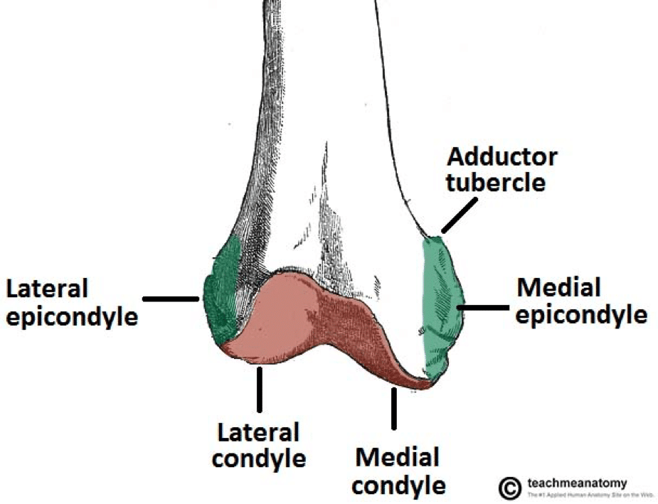
The medial condyle of femur (medial femoral condyle, internal condyle of femur, or tibial condyle of femur) is the medially located, round, articular eminence on the distal end of the femur. It is one of the two articular condyles of the femur, the other being the lateral condyle. It contains the medial epicondyle of femur and adductor tubercle.

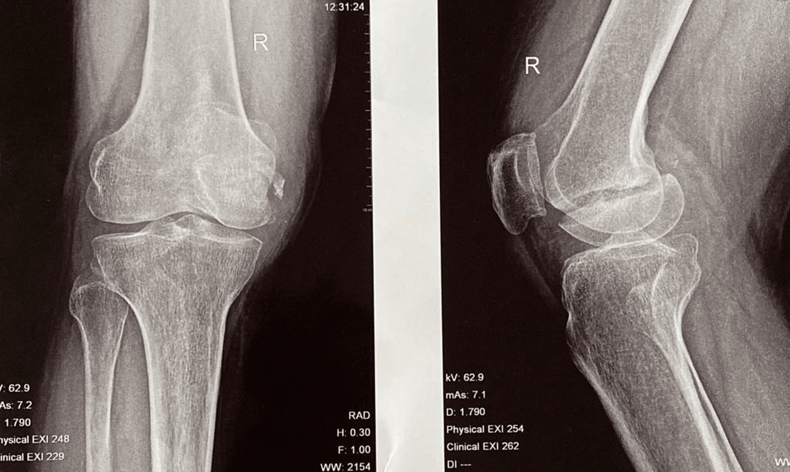
A case of compression fracture of medial tibial plateau and medial femoral condyle combined with
The distal femur is the area of the leg just above the knee joint. Distal femur fractures most often occur either: In older people whose bones are weak, or. In younger people who have high energy injuries, such as from a high-speed motor vehicle collision. In both the elderly and the young, the breaks may extend into the knee joint and, in some.
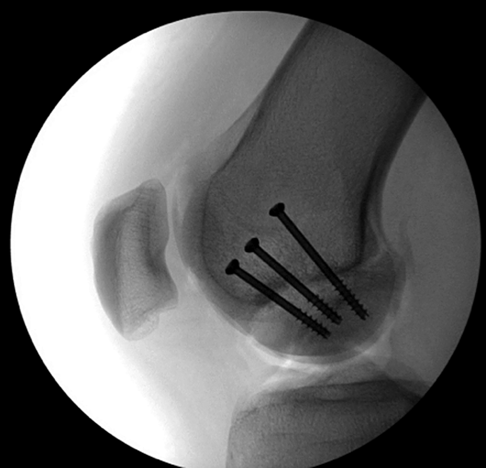
A Novel Technique for Fixation of a Medial Femoral Condyle Fracture using a Calcaneal Plate
Distal femur fractures include fractures of the supracondylar and intercondylar region and are relatively common injuries. The goals of treatment follow AO principles of anatomic reduction of the articular surface, restoration of limb alignment, length, and rotation. Despite improvements in implant design, management of distal femur fractures remains a challenge; fractures are often comminuted.

Early femoral condyle insufficiency fractures after total knee arthroplasty treatment with
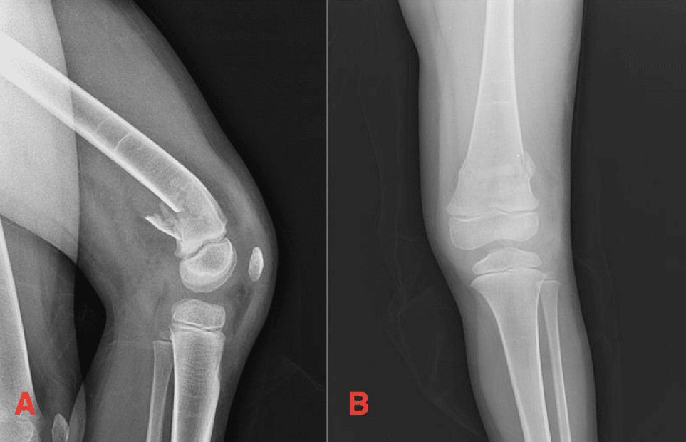
Summary. Medial condyle fractures are a rare traumatic injury most commonly occurring in children 2-9 years of age caused by a fall onto an elbow. Diagnosis is typically made with plain films, although these injuries can be difficult to detect, especially in young children. MRI may be of utility in assisting with prompt diagnosis.

Figure 1 from Hoffa's fracture of the medial femoral condyle in a child treated with open
The medial condyle is one of the two projections on the lower extremity of femur, the other being the lateral condyle . The medial condyle is larger than the lateral (outer) condyle due to more weight bearing caused by the centre of mass being medial to the knee. On the posterior surface of the condyle the linea aspera (a ridge with two lips.
Medial Femoral Condyle Fracture
Femoral medial condyle fracture is a rare fracture. As with any articular injury, anatomical restoration of the joint surface must be obtained, then lag screw fixation is required. In fractures with a vertical fracture line, a buttress plate is necessary to counteract the vertical shear forces. Nevertheless, there are no available anatomical.
Cureus A Case of Distal Femur Medial Condyle Hoffa Type II(C) Fracture Treated with Headless
Distal femur fractures are traumatic injuries involving the region extending from the distal metaphyseal-diaphyseal junction to the articular surface of the femoral condyles. Diagnosis is made radiographically with CT studies often required to assess for intra-articular extension. Treatment is generally operative with ORIF, intramedullary nail.

Distal Femur Fractures Trauma Orthobullets
A femoral condyle is the ball-shape located at the end of the femur (thigh bone). There are two condyles on each leg known as the medial and lateral femoral condyles. If there is a fracture (break) in part of the condyle, this is known as a fracture of the femoral condyle. Physiotherapy is very important during the rehabilitation following a.
Medial Femoral Condyle Fracture
Distal femoral fractures involve the femoral condyles and the metaphyseal region and are often the result of high energy trauma such as motor vehicle accidents or a fall from a height. In the elderly, they may occur as a domestic accident 1-3. ICD-11 NC72.6Z.
- Restaurants At Eagle Street Pier
- What Beers Are Gluten Free Australia
- What Time Is In Laos
- Academy Of Early Education Sydenham
- Laws About Domestic Violence In Australia
- Paradise Lost On The Burnett
- Why Does Pubic Hair Grow So Fast
- Japanese Maneki Neko Lucky Cat
- Oh Happy Day Oh Happy Day Lyrics
- Temperature In Perth In May